Lucas M. Tomatis1, Rita L. O. Pastore1, Joseph Torres Guerrero1, Gabriela L. Sidra1, Martín Jaume2, Luciana Pérez Mora4, Laura Svidler López1
1 Division of Surgery, Colorectal Surgery Section
2 Division of Infectious Diseases
3 Division of Pathology
Hospital General de Agudos Juan A. Fernández, Buenos Aires, Argentina
Correspondence
Laura Svidler López lausvidlerlopez@gmail.com
Received: April 20, 2026
Accepted: July 20, 2026
How to cite: Tomatis LM, Pastore RLO, Torres Guerrero J, Sidra GL, Jaume M, Pérez Mora L, Svidler López L. Abdominal Tuberculosis: Diagnostic Challenges and Clinical Implications in a Tertiary Referral Hospital in Buenos Aires, Argentina. Rev. argent. coloproctología 2026;37(3):.doi: 10.46768/jd7bf717
ABSTRACT
Background: Abdominal tuberculosis (ATB) is an uncommon extrapulmonary manifestation with a nonspecific presentation, which frequently delays diagnosis. Evidence in adult populations in Argentina remains limited.
Objective: To describe clinical features, diagnostic delay, surgical requirements, and outcomes in patients with ATB, and to compare findings according to HIV status.
Methods: We conducted a retrospective observational study of adult patients diagnosed with ATB between January 2017 and October 2025 at Hospital Juan A. Fernández. Diagnosis was confirmed by microbiological, molecular (Xpert® MTB/RIF), and/or histopathological methods.
Results: Twelve patients were included (median age 34 years, IQR, 27–39); 58,3% were female and 58,3% were HIV-positive. The most common symptoms were abdominal pain (91,7%), anemia (83,3%), weight loss (67,7%), and fever (67,7%). Disease distribution was intestinal in 41,7%, anorectal in 41,7%, and peritoneal in 33,3%, with disseminated disease in 83,3%. A presumptive diagnosis of ATB was established in 50% of patients, whereas the remainder were initially misdiagnosed as acute inflammatory abdomen or malignancy. Overall, 58,3% required surgical intervention. An elevated postoperative complication rate (57.1%) was observed, with the need for reoperation in three patients.
Diagnosis was confirmed by histopathology (100%), culture (91,7%), and Xpert® MTB/RIF (50%), with most patients having more than one positive diagnostic modality. The median diagnostic delay was 32 days (IQR, 21–44). HIV-negative patients required surgery more frequently than HIV-positive patients (100% vs. 28,6%; p = 0.03).
Conclusions: Abdominal tuberculosis should be considered in the differential diagnosis of acute abdomen and anorectal disease in high-prevalence settings, particularly in patients with HIV coinfection. In this series, anorectal involvement was frequent, and HIV-positive patients required less surgical intervention, which may reflect a higher level of clinical suspicion and earlier diagnosis. The disease is associated with diagnostic delays and may require surgical management in complicated cases and, to a lesser extent, for diagnostic purposes. Postoperative outcomes may be complex, with a higher rate of complications. A high index of clinical suspicion and a multimodal, interdisciplinary approach, including rapid molecular methods, are essential to reduce diagnostic delays and improve outcomes.
Keywords: Abdominal tuberculosis; Intestinal tuberculosis; Anorectal tuberculosis; Peritoneal tuberculosis; HIV; Diagnosis; Diagnostic delay
Tuberculosis (TB) remains a major global public health problem, particularly in low- and middle-income countries. In Argentina, an increase in the number of cases has been observed in recent years, of which approximately 11.9% correspond to extrapulmonary forms.1,2 Abdominal tuberculosis (ATB) accounts for approximately 11% of peritoneum, lymph nodes, and solid organs. Clinical manifestations are often nonspecific and may mimic malignancy or inflammatory bowel disease, frequently leading to delays in diagnosis.3,4
The paucibacillary nature of the disease and the low sensitivity of diagnostic tests often require invasive procedures for tissue acquisition.5,6 The Xpert® MTB/RIF assay, based on real-time PCR, allows detection of Mycobacterium tuberculosis complex DNA and rifampin resistance in approximately two hours, although its availability remains limited in some centers. In contrast, mycobacterial culture may take three to four weeks, further contributing to diagnostic delay.7
Despite its clinical relevance, published data on ATB in the adult population in Argentina are scarce. In the largest available series, Vasen et al.⁸ (2016) highlighted the nonspecific presentation of the disease and reported an overall mortality of 8%, which was higher among patients with HIV coinfection, underscoring the need to update the available evidence in Argentina.
The aim of this study was to describe the clinical characteristics, anatomic distribution, diagnostic delay, management, and outcomes of patients with ATB at a tertiary referral center in the City of Buenos Aires, and to compare findings between immunocompetent patients and those infected with HIV.
A retrospective observational study was conducted that included adult patients (≥18 years) diagnosed with ATB between January 2017 and October 2025 at Hospital Juan A. Fernández, Diagnosis was established based on microbiologic tests (Ziehl–Neelsen staining and culture), molecular testing (Xpert® MTB/RIF), and/or compatible histopathologic findings.
Data collected included demographics, clinical presentation, anatomic distribution, HIV status, imaging findings, presumptive diagnosis before ATB confirmation, diagnostic delay (time interval in days between symptoms onset and diagnostic confirmation using microbiological, molecular, and/or histopathological methods), surgical management, complications, and outcomes.
.
Statistical Analysis
Continuous variables are presented as the median and interquartile range (IQR) and were compared using the Mann–Whitney U test, performed with JASP version 0.97.1 (JASP Team, University of Amsterdam, Amsterdam, the Netherlands). Categorical variables are presented as frequencies and percentages and were compared using Fisher's exact test, performed with the GraphPad QuickCalcs calculator (GraphPad Software, San Diego, CA, USA). All statistical tests were two-sided, and a P value < 0.05 was considered statistically significant.
Twelve patients were included, of whom 7 (58.3%) were female. The median age was 34 years (IQR, 27–39). HIV infection was present in 58.3% of patients, and 50% had concomitant pulmonary TB. Patient characteristics, clinical presentation, and anatomic distribution are summarized in Table 1.
Abdominal pain was the most common symptom (91.7%), followed by weight loss and fever (66.7% each).
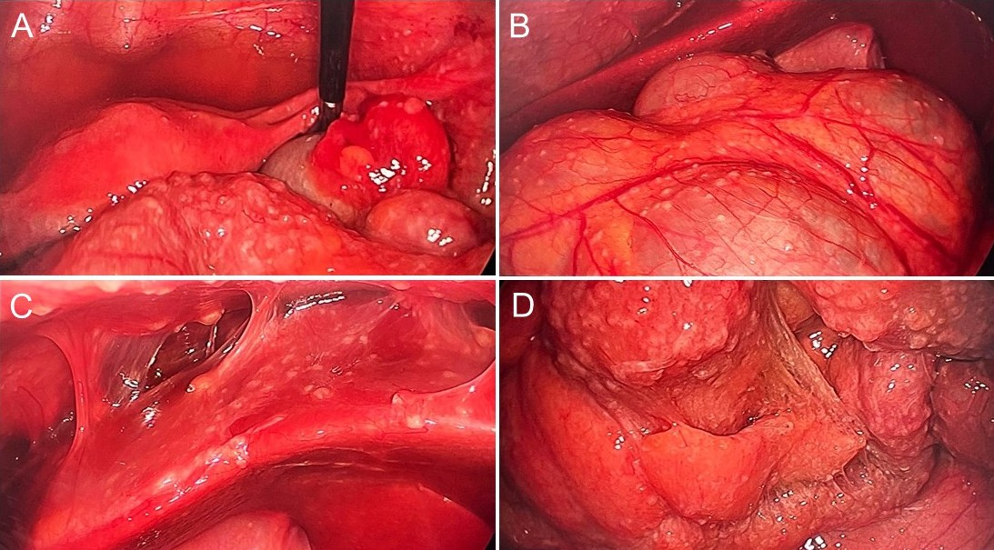
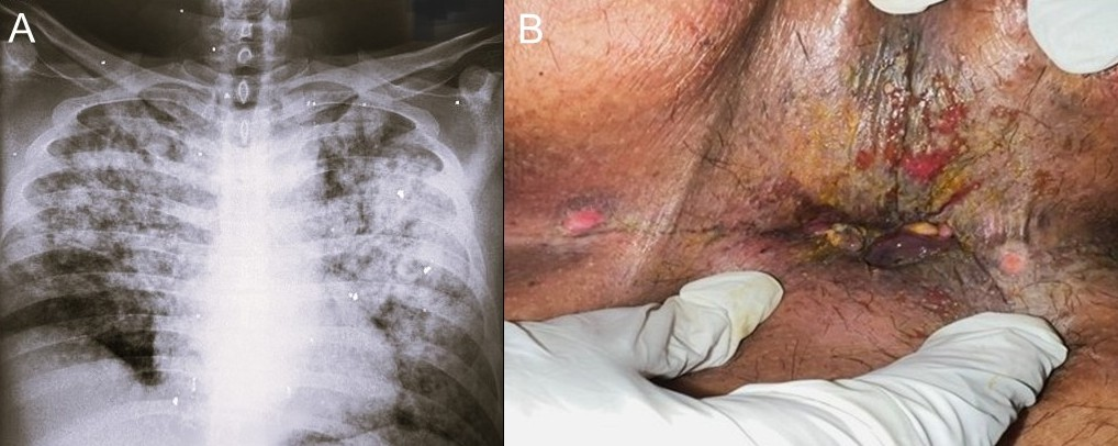
Intestinal and anorectal involvement were observed in 41.7% each, and peritoneal involvement was present in 33.3% (Fig. 1). Disseminated disease occurred in 83.3% of patients, including multifocal abdominal involvement (n = 4) and/or concomitant or prior pulmonary TB (n = 7).
Ano-perianal disease (including abscesses, fistulas, and ulcerations) was identified in 4 patients; of these, 3 were HIV-positive and had active pulmonary TB (Fig. 2).
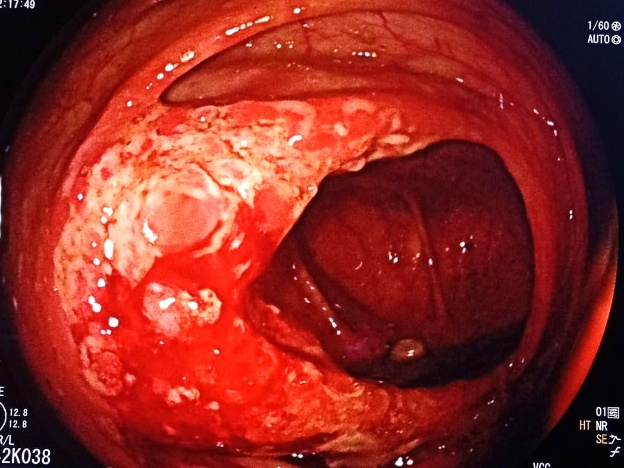
Rectal involvement presented as ulcerated pseudotumoral lesions in 3 HIV-positive patients, 2 of whom had active pulmonary TB.
Anemia and hypoalbuminemia were present in 83.3% of patients, and leukopenia in 41.7%. In HIV-positive patients, the median CD4 count was 313 cells/mm³ (IQR, 204–683); 3 (42.8%) patients had CD4 counts <200 cells/mm³, and 2 had detectable viral load.
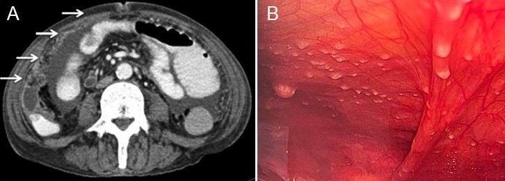
All patients underwent imaging evaluation. Computed tomography (CT) of the chest, abdomen, and pelvis was performed in 10 patients, and abdominal ultrasound in 2. Findings suggestive of abdominal TB included ileocecal wall thickening (Fig. 3), stenotic ceco-ascending lesions, mesenteric lymphadenopathy, ascites (free and loculated), miliary peritoneal nodules (Fig. 4), bowel loop conglomerates, and omental/peritoneal thickening (“omental cake”).

In the three patients with rectal involvement, colonoscopy with biopsy was performed. One of them also had associated ileocecal TB (Fig. 5).
Xpert® MTB/RIF testing was performed in 6 patients (50%), using pleural fluid, ascitic fluid, perianal discharge, endoscopic biopsies, or surgical specimens.
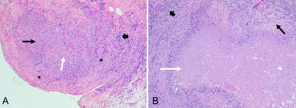
Mycobacterial culture was positive in all 11 tested patients. Histopathology confirmed the diagnosis in the remaining patient with a rectal lesion (Fig. 6). Ziehl–Neelsen staining was positive in 45% of culture-positive cases.
A presumptive diagnosis of ATB was made in 6 patients (50%). Initial misdiagnoses included acute appendicitis (n = 2), inflammatory acute abdomen (n = 1), peritoneal carcinomatosis (colonic or ovarian origin) (n = 2), and rectal cancer (n = 1).
Surgery was required in 7 patients (58.3%), mainly due to inflammatory acute abdomen (n = 3), perforation (n = 1), obstruction (n = 1), or diagnostic uncertainty (n = 2). Four patients (57.1%) experienced postoperative complications, and three required reoperation. Presumptive diagnoses, surgical findings and outcomes are summarized in Table 2.
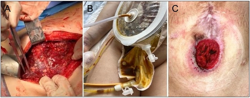
The median diagnostic delay was 32 days (IQR, 21–44), ranging from 15 to 154 days. The longest delay occurred in a patient with a 4-month history of abdominal pain, weight loss, diarrhea, and fever. CT findings suggested ATB; however, diagnostic laparotomy revealed severe fibroadhesive peritonitis with sealed perforation (Fig. 7).
| Variables | Patients N = 12 |
|---|---|
| Age (years), median (IQR) | 34 (27–39) |
| Female sex, n (%) | 7 (58.3) |
| HIV coinfection, n (%) | 7 (58.3) |
| Pulmonary TB, n (%) Concomitant Prior | 6 (50.0) 1 (8.3) |
| Symptoms, n (%) Abdominal pain Weight loss Fever Cough Night sweats Anal pain Nausea/vomiting Dyspnea Diarrhea Hematochezia | 11 (91.7) 8 (66.7) 8 (66.7) 7 (58.3) 5 (41.7) 5 (41.7) 4 (33.3) 4 (33.3) 2 (16.7) 2 (16.7) |
| Anatomical location, n (%) Ileocecal Anorectal Peritoneal Small intestine Multifocal | 5 (41.7) 5 (41.7) 4 (33.3) 1 (8.3) 4 (33.3) |
IQR = interquartile range. TB = tuberculosis.
Comparative findings between immunocompetent and HIV-infected patients are presented in Table 3. The need for surgical intervention was significantly higher among immunocompetent patients (100% vs 28.6%; p = 0.03). A higher proportion of disseminated tuberculosis and greater anorectal involvement were observed in the HIV-infected group, whereas peritoneal TB predominated among HIV-negative patients; however, these differences did not reach statistical significance.
| Presumptive diagnosis | HIV | Intraoperative finding / Procedure| | Outcome |
|---|---|---|---|
| Acute appendicitis* | Negative | Appendicitis → laparoscopic appendectomy | No complications |
| Acute appendicitis | Positive | Appendicitis → laparoscopic appendectomy | ECF, retroperitoneal abscess, adnexitis → right colectomy, ileocolic anastomosis, adnexectomy |
| Perforated acute abdomen, palpable RLQ mass | Negative | Fibroadhesive peritonitis, miliary nodules, ileal perforation (not exteriorizable) → exploratory laparotomy, omental biopsy, Foley catheter drainage | Leak → re-laparotomy, VAC, open abdomen → ECF → VAC |
| Ascites, ovarian irregularity, ↑CA-125. Carcinomatosis vs ATB | Negative | Diffuse peritoneal thickening, omental nodules, ascites → exploratory laparoscopy, biopsy, fluid sampling | Persistent fever → percutaneous drainage |
| Inflammatory acute abdomen† | Negative | Cecal thickening, mesenteric lymphadenopathy, peritoneal nodules, loculated ascites → exploratory laparoscopy, biopsies, fluid sampling | No complications |
| Obstructive colon cancer,palpable RLQ mass, carcinomatosis, pulmonary metastases⁋ | Positive | Ascending colon stenotic lesion, peritoneal nodules, ascites → exploratory laparotomy, biopsies, fluid sampling, double-barrel ileostomy | Ileostomy necrosis → re-ileostomy |
| Disseminated TB: (nodal, abdominal, pulmonary, pleural); thoracic / ascitic studies negative | Negative | Diffuse whitish nodules, mesenteric lymphadenopathy, ascites → exploratory laparoscopy, lymph node biopsy, fluid sampling | No complications |
ECF = enterocutaneous fistula. RLQ = right lower quadrant. VAC = vaccum assisted closure. ATB = abdominal tuberculosis.
* Active pulmonary TB under antituberculous treatment
† Father deceased due to TB
⁋ Postoperative confirmation of miliary pulmonary TB after abdominal surgery
| Variable | HIV negative (N = 5) | HIV positive (N = 7) | p |
|---|---|---|---|
| Age (years), median (IQR) | 33 (25–37) | 34 (30–43) | 0,33* |
| Female sex, n (%) | 3 (60,0) | 4 (57,1) | 1,00† |
| Concurrent/prior pulmonary TB, n (%) | 2 (40,0) | 5 (71,4) | 0,66† |
| Symptoms, n (%) Intestinal/anorectal Constitutional | 5 (100) 5 (100) | 7 (100) 5 (71,4) | — 1,00† |
| Site of involvement, n (%) Intestinal/visceral Peritoneal Anorectal Disseminated disease | 1 (20,0) 3 (60,0) 1 (20,0) 3 (60,0) | 4 (57,1) 1 (14,3) 4 (57,1%) 7 (100%) | 0,29† 0,22† 0,29† 0.15† |
| Diagnostic delay (days), median (IQR) | 18 (8–97) | 30 (14–34) | 0,87* |
| Need for surgical intervention, n (%) | 5 (100) | 2 (28,6) | 0,03 † |
IQR = interquartile range. TB = tuberculosis.
* Mann–Whitney U test. † Fisher’s exact test. p < 0.05 was considered statistically significant.
This case series highlights the persistence of abdominal tuberculosis (ATB) as a relevant public health problem in the urban setting of Buenos Aires, particularly among vulnerable populations. Abdominal pain, present in more than 90% of cases, is a nonspecific symptom shared by multiple conditions. This was reflected in the wide range of initial diagnoses, including acute appendicitis, peritoneal carcinomatosis, and rectal cancer, consistent with prior reports.9,10 The presence of constitutional symptoms (fever, weight loss) and epidemiologic context (immunosuppression, TB exposure) are key factors in guiding clinical suspicion.
Consistent with previous reports, ileocecal lymph node involvement was the most common site of disease.9,11 Likewise, peritoneal involvement was a frequent finding, consistent with the results reported by Moolla et al.¹⁰ in a South African cohort and by Vasen et al.⁸ in an Argentinian case series. Peritoneal TB accounts for a substantial proportion of ATB (25–50% in patient series)11,12 and presents in three main pathologic forms that may coexist: wet or ascitic
(free or loculated ascites), fibrotic-fixed (peritoneal thickening and adhesions), and dry or plastic (nodular peritoneal thickening and omental involvement (“omental cake”).11,13 Peritoneal TB may clinically and radiologically mimic peritoneal carcinomatosis of colorectal or ovarian origin, including nonspecific elevation of CA-125 due to mesothelial inflammation.11,14–16 Factors such as younger age, prior TB, or HIV coinfection may suggest an infectious etiology, and laparoscopy with multiple biopsies remains the gold standard for diagnostic confirmation in these cases.11,14,17
A distinctive finding of our series was the high proportion of anorectal involvement (41,7%), in contrast to the literature, where it represents less than 1% of gastrointestinal TB cases.11,12 This difference may reflect both population-specific epidemiologic characteristics and increased detection in a specialized proctology setting. Anorectal TB presents a broad clinical spectrum, including recurrent fistulas, abscesses, superficial ulcers with necrotic bases and nonindurated edges, strictures, and submucosal tumor-like lesions, often overlapping with inflammatory bowel disease and malignancy.11–18
In our cohort, it predominated in male patients, with a high rate of HIV coinfection and frequent association with concomitant pulmonary TB.
Anal fistula is the most common presentation (80%–91%) and lacks distinguishing features compared with cryptoglandular disease.12 Sultan et al.19 described seven cases of recurrent perianal fistulas/abscesses in male patients treated with combined surgery and antituberculous therapy, achieving complete resolution without recurrence, and noting that HIV coinfection was not associated with increased incidence.
Rectal TB is rare, with only 24 cases reported up to 2022 according to a systematic review,20 and is usually associated with active pulmonary TB. Converserly, series such as that of Tago et al.13 show that up to 42% of perianal cases occur without pulmonary involvement and are associated with significantly longer disease duration (66.4 vs 8.3 months) due to diagnostic delay. Consistent with this, in our cohort, patients without pulmonary TB were initially suspected of having neoplastic disease. Although the absence of respiratory symptoms may reduce clinical suspicion, TB should be considered in chronic, recurrent, or nonhealing anorectal lesions.18,20 The tuberculin skin test may be useful, with positivity reported in approximately 75% of perianal TB cases in immunocompetent patients.18
Treatment of rectal TB is primarily medical, based on standard 6-month antituberculous regimens, with high cure rates; surgery is reserved for cases with complications, lack of response, or diagnostic uncertainty.20 In anorectal TB, although some fistulas may resolve with medical therapy alone, combined surgical management is recommended for adequate control of anorectal sepsis.18,19
The mean diagnostic delay was 32 days, within the range reported in other series, although with wide variability, reaching 154 days in a complex case.8 This extreme case demonstrates the consequences of delayed diagnosis, including local progression with intestinal perforation, clinical deterioration, and significant surgical morbidity (chronic enterocutaneous fistula).
Because up to 75% of extrapulmonary TB cases have negative smear results and cultures require weeks,11 Xpert® MTB/RIF was a key tool for rapid diagnosis in most cases. However, its sensitivity in paucibacillary extrapulmonary samples is variable,7 necessitating a multimodal approach that includes histopathology, culture, and, when required, invasive procedures to obtain adequate tissue samples.6 This limitation is illustrated by a case of disseminated TB in which Xpert® MTB/RIF was positive only in a laparoscopic lymph node biopsy, with negative results in ascitic and pleural fluid.
The rate of surgical intervention in our cohort was within the 30%–60% range reported for ATB3,8 and was mainly associated with complications such as bowel obstruction and acute inflammatory abdomen. These findings underscore the importance of including TB in the differential diagnosis of acute abdomen, particularly in high-prevalence settings and in patients with HIV, as well as ensuring adequate tissue sampling during surgery.21
A high rate of postoperative complications (57.1%) was observed, and three patients required reoperation. This could be linked to delayed diagnosis and the general and local deterioration associated with the disease.
The prevalence of HIV coinfection (58,3%) was markedly higher than the 13% reported in other ATB series,8 likely reflecting the role of our institution as a referral center for immunocompromised patients. This is clinically relevant, as advanced immunosuppression is associated with an increased risk of disseminated and multifocal extrapulmonary disease.22
Comparative analysis did not demonstrate significant differences in clinical presentation or diagnostic delay between patients with and without HIV, although a nonsignificant trend toward more disseminated disease was observed in the HIV group. The only significant difference was a higher need for abdominal surgery among immunocompetent patients. This finding may be explained by lower initial clinical suspicion of TB in this group, leading to misclassification as acute surgical or neoplastic disease,8,12,14,15 whereas TB is more readily considered in immunocompromised patients, facilitating a less invasive diagnostic approach.22
This study has several limitations, including its retrospective design and small sample size, which may overestimate the magnitude of the observed differences when results are expressed as percentages. Accordingly, the findings should be interpreted with caution, as the limited statistical power may have reduced the ability to detect differences between groups.
Abdominal tuberculosis is a challenging diagnosis due to its nonspecific presentation and its frequent mimicry of inflammatory and neoplastic conditions. In settings with high prevalence of tuberculosis and HIV coinfection, it should be included in the differential diagnosis of acute abdomen, as well as in patients presenting with persistent abdominal pain, ascites, abdominal masses, and chronic, recurrent, or atypical anorectal disease, particularly in immunocompromised individuals. The observed diagnostic delay highlights the importance of maintaining a high level of suspicion, while the incorporation of rapid molecular methods may help shorten diagnostic time. Surgery is a key component of both diagnosis and treatment in complicated cases, and the surgical course can be complex in patients with late-stage or advanced disease, emphasising the significance of timely diagnosis.
This series underscores the high frequency of anorectal involvement and the elevated rate of HIV coinfection, with a lower requirement for surgical intervention in this group, possibly reflecting earlier clinical suspicion and diagnosis. A multidisciplinary and multimodal approach is essential to optimize clinical suspicion, reduce diagnostic delays, and improve outcomes.
Author Contributions:
LMT: Data curation, Research, Writing – original draft.
RLOP: Conceptualization, Methodology, Formal analysis, Writing – original draft, Writing – review and editing, Supervision.
JTG: Data curation, Research, Writing – original draft.
GLS: Research, Writing – original draft.
MJ: Research, Writing – original draft.
LPM: Research, Writing – original draft.
LSL: Conceptualization, Methodology, Research, Writing – original draft, Writing – review and editing, Supervision.
Conflict of interest statement: None.
Funding: None.
Data availability statement: The data are publicly available.
ORCIDs:
Lucas Tomatis: https://orcid.org/0000-0002-4593-3002
Rita Pastore: https://orcid.org/0000-0001-5240-9068
Gabriela Sidra: https://orcid.org/0000-0002-0651-6353
Joseph Torres: https://orcid.org/0009-0005-2624-7428
Martin Jaume: https://orcid.org/0009-0004-3205-3681
Luciana Pérez Mora: https://orcid.org/0009-0005-1858-5443
Laura Svidler López: https://orcid.org/0000-0001-5898-6667
Ministerio de Salud de la Nación. Boletín Epidemiológico N° 8: Notificación de casos de tuberculosis en Argentina. Buenos Aires: Ministerio de Salud de la Nación; 2025.
Ministerio de Salud de la Nación. Boletín Epidemiológico Nacional. Semana Epidemiológica N° 53. Buenos Aires: Ministerio de Salud de la Nación; 2026.
Al Zanbagi AB. Gastrointestinal tuberculosis: a systematic review of epidemiology, presentation, diagnosis and treatment. World J Clin Cases. 2021;12(7):1290–1305.
Sampath A, Mani S. Diagnostic evaluation and management of abdominal tuberculosis. Indian J Tuberc. 2025;72(Suppl 1):S7–S11.
Jha DK, Menon Pathiyil M, Sharma V. Evidence based approach to diagnosis and management of abdominal tuberculosis. Indian J Gastroenterol. 2023;42(1):17–31.
Tahiri M, Goh KL, Abbas Z, Epstein D, Chen M-H, Mulder CJJ, Puri AS, Schultz M, LeMair A, Melberg J. Digestive Tract Tuberculosis Guideline. J Clin Gastroenterol. 2023;57(7):643-650. doi:10.1097/MCG.0000000000001819.
Kohli M, Schiller I, Dendukuri N, Yao M, Dheda K, Denkinger CM, Schumacher SG, Steingart KR. Xpert MTB/RIF Ultra and Xpert MTB/RIF assays for extrapulmonary tuberculosis and rifampicin resistance in adults. Cochrane Database Syst Rev. 2021;1(1):CD012768. doi:10.1002/14651858.CD012768.pub3.
Vasen W, Mauriño E, Ferro D, Brotto C, Fernández Marty P, Cabanne A. Síndrome de tuberculosis abdominal: análisis de 100 casos clínicos. Acta Gastroenterol Latinoam. 2016;46:205–12.
Tobin EH, Khatri AM. Abdominal Tuberculosis. In: StatPearls [Internet]. Treasure Island (FL): StatPearls Publishing; 2026 Jan–. Updated 2025 Feb 6. Bookshelf ID: NBK556115. PMID: 32310575. Available from: https://www.ncbi.nlm.nih.gov/books/NBK556115/
Moolla MS, Abdul Karim SS. Abdominal tuberculosis in South Africa: clinical features and diagnostic challenges. S Afr Med J. 2011;101:745–8.
Alsaeed M, Alghamdi A, Aldawish A, et al. Clinical Characteristics and Outcomes of Gastrointestinal Tuberculosis in Saudi Arabia: A 10-year Retrospective Cohort Study. Int J Mycobacteriol. 2026;15(1):38-44. doi:10.4103/ijmy.ijmy_13_26
Eraksoy H. Gastrointestinal and Abdominal Tuberculosis. Gastroenterol Clin North Am. 2021;50(2):341-360. doi:10.1016/j.gtc.2021.02.004
Tago S, Hirai Y, Ainoda Y, Fujita T, Takamori M, Kikuchi K. Perianal tuberculosis: A case report and review of the literature. World J Clin Cases. 2015;3(9):848-52. doi: 10.12998/wjcc.v3.i9.848.
Ladumor H, Al-Mohannadi S, Ameerudeen FS, Ladumor S, Fadl S. TB or not TB: a comprehensive review of imaging manifestations of abdominal tuberculosis and its mimics in adults. Clin Imaging. 2021;76:130-43.
Cho JH, Kim SS. Peritoneal carcinomatosis and its mimics: review of ct findings for differential diagnosis. J Belg Soc Radiol. 2020;104(1):8. doi: 10.5334/jbsr.1940.
Ozan H, Ozerkan K, Orhan A. Peritoneal tuberculosis mimicking peritoneal carcinomatosis. Eur J Gynaecol Oncol. 2009;30:645–8.
Sohail AH, Khan MS, Sajan A, Williams CE, Amodu L, Hakmi H, et al. Diagnostic accuracy of CT in differentiating peritoneal tuberculosis from carcinomatosis. Clin Imaging. 2022;82:198–203. doi: 10.1016/j.clinimag.2021.11.023.
Gupta PJ. Ano-perianal tuberculosis – solving a clinical dilemma. Afr Health Sci. 2005;5(4):345–47.
Sultan S, Azria F, Bauer P, Abdelnour M, Atienza P. Anoperineal tuberculosis: diagnostic and management considerations in seven cases. Dis Colon Rectum. 2002;45:407–10.
Chaudhary P, Nagpal A, Padala SB, Mukund M, Bansal LK, Lal R. Rectal tuberculosis: A systematic review. Indian J Tuberc. 2022;69(3):315–22.
Debi U, Ravisankar V, Prasad KK, Sinha SK, Sharma AK. Abdominal tuberculosis of the gastrointestinal tract: revisited. World J Gastroenterol. 2014;20:14831–40.
Swaminathan S, Padmapriyadarsini C, Narendran G. HIV-associated tuberculosis: clinical update. Clin Infect Dis. 2010;50(10):1377-86. doi: 10.1086/652147.