Andrea V. Ruchinsky1, Dana Santos2, Florencia Litovska3, Gabriela Rio Gorrini3
1General Surgery Resident, General Surgery Department, Hospital Carlos G. Durand, Ciudad de Buenos Aires, Argentina
2General Surgery Resident, General Surgery Department, Hospital Aeronáutico Central, Ciudad de Buenos Aires, Argentina
3Colorectal Surgeon, General Surgery Department, Coloproctology Division, Hospital Carlos G. Durand, Ciudad de Buenos Aires, Argentina
Correspondence
Andrea V. Ruchinsky andrea-v99@hotmail.com
Received: February 25, 2026
Accepted: June 16, 2026
How to cite: Ruchinsky AV, Santos D, Litovska F, Rio Gorrini G. Primary extramedullary plasmacytoma of the ascending colon. Rev. argent. coloproctología 2026;37(3). doi: 10.46768/010c1d38
ABSTRACT
Solitary extramedullary plasmacytoma is a rare plasma cell neoplasm, and colonic involvement is exceedingly uncommon. We report the case of a 57-year-old man diagnosed with a right colonic plasmacytoma during the evaluation of anemia identified in the preoperative workup for elective cholecystectomy. Further investigation revealed a localized lesion without evidence of systemic involvement or criteria for multiple myeloma. The patient underwent laparoscopic right hemicolectomy with complete tumor resection and an uneventful postoperative recovery. Histopathological examination and immunohistochemical staining confirmed the diagnosis of solitary extramedullary plasmacytoma. This case highlights the importance of thorough evaluation of unexplained anemia and underscores the role of complete surgical excision as a curative approach for localized colonic plasmacytoma.
Keywords: solitary extramedullary plasmacytoma; colonic plasmacytoma; plasma cell neoplasms; multiple myeloma; immunohistochemistry; laparoscopic right hemicolectomy.
Solitary extramedullary plasmacytoma (SEP) is a rare plasma cell neoplasm characterized by a localized monoclonal proliferation in soft tissues, in the absence of bone marrow involvement and without meeting the diagnostic criteria for multiple myeloma.¹ Together with solitary bone plasmacytoma, SEP forms part of the spectrum of plasma cell neoplasms, distinguished primarily by its anatomic location and biological behavior. It accounts for approximately 3% of all plasma cell neoplasms, while involvement of the lower gastrointestinal tract is exceedingly rare, with only a limited number of cases reported in the literature.² Accurate recognition and diagnostic characterization are essential, as the prognosis, treatment approach, and follow-up strategy differ substantially from those of multiple myeloma. Although SEP generally follows a more favorable clinical course, it carries a risk of progression to multiple myeloma, underscoring the importance of comprehensive initial staging and long-term clinical surveillance.
A 57-year-old man undergoing preoperative evaluation for elective cholecystectomy was found to have moderate anemia. Hematologic evaluation revealed positive direct and indirect Coombs tests and positive panagglutinins.
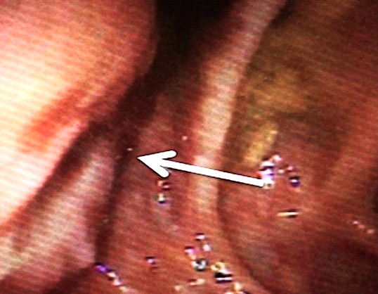
Colonoscopy demonstrated a raised, centrally ulcerated lesion in the ascending colon. Multiple biopsy specimens were obtained from the lesion (Fig. 1).
Histopathologic examination demonstrated diffuse infiltration by mature plasma cells and scattered B lymphocytes. Immunohistochemical staining showed positivity for plasma cells (CD138) and kappa light chains, with negativity for lambda light chains, findings consistent with plasmacytoma.
Positron emission tomography/computed tomography (PET/CT) demonstrated hypermetabolic mural thickening of the ascending colon near the hepatic flexure (SUVmax, 8.4), with adjacent hypermetabolic lymphadenopathy suspicious for metastatic involvement.
The diagnostic criteria for solitary plasmacytoma established by the International Myeloma Working Group and the Argentine Society of Hematology were assessed to exclude multiple myeloma, including: 1) a soft-tissue mass composed of clonal plasma cells; 2) serum monoclonal gammopathy and/or low concentrations or absence of urinary light chains; 3) absence of clonal plasma cells on bone marrow biopsy; and 4) absence of myeloma-related end-Laboratory testing showed elevated serum free kappa light chains (150 mg/L) and lambda light chains (12.69 mg/L), with weakly positive urinary Bence Jones protein of the kappa type. Bone marrow biopsy was normal. There was no hypercalcemia, renal impairment, or anemia. Whole-body CT showed no osteolytic lesions.
A presumptive diagnosis of SEP of the ascending colon was made. Following review by a multidisciplinary team, comprising colorectal surgeons, oncologists, hematologists, pathologists, and radiologists, laparoscopic right colectomy with primary ileocolic anastomosis was performed for local disease control and definitive histopathologic diagnosis. The procedure was completed without intraoperative complications.
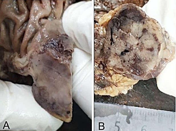
Gross examination revealed a firm, whitish lesion measuring 2.8 × 2.5 cm in the ascending colon, with slightly raised borders and central ulceration. The lesion extended through the full thickness of the colonic wall into the pericolic fat and caused approximately 60% luminal stenosis (Fig. 2A). Within the pericolic adipose tissue, rounded nodes measuring up to 2.9 cm were identified, corresponding grossly to a nodal conglomerate (Fig. 2B).
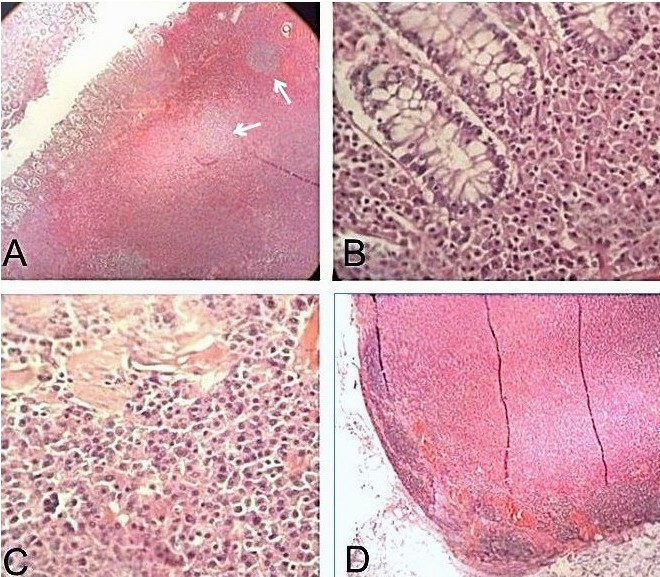
Microscopic examination revealed a diffuse proliferation of atypical plasmacytoid cells measuring 2.8 × 2.5 cm, with a predominantly interstitial growth pattern and displacement of the colonic glands. The neoplastic infiltrate extended from the mucosa, where focal superficial erosion was present, through the subserosal fibro-adipose tissue (Fig. 3A–C). Fifteen regional lymph nodes were identified, 3 of which were involved by the neoplasm and corresponded to the previously identified nodal conglomerate (Fig. 3D). All surgical margins were negative for neoplasia.
Immunohistochemical studies demonstrated strong, diffuse CD138 expression by the neoplastic cells, with kappa light-chain restriction, supporting a clonal plasma cell population. Only rare CD20-positive B cells and CD3-positive T cells were identified, scattered throughout the stromal background. Taken together, the morphologic and immunophenotypic findings were consistent with a plasma cell neoplasm and confirmed the diagnosis of extramedullary plasmacytoma.
The postoperative course was uneventful, with no complications. The patient was discharged on postoperative day 5, with outpatient follow-up by the colorectal surgery and hematology services.
The diagnosis of SEP requires integration of clinical, radiologic, histopathologic, and immunohistochemical findings, together with comprehensive systemic staging to exclude multiple myeloma, as this distinction has important implications for treatment, prognosis, and follow-up. Given the rarity of colorectal involvement, the available evidence is largely limited to case reports and small case series; therefore, the optimal diagnostic and therapeutic approach remains uncertain.
Clinically, patients may present with abdominal pain, changes in bowel habits, gastrointestinal bleeding, or anemia; however, many are asymptomatic, with the diagnosis made incidentally during evaluation for other indications, as in the present case.3,4
Definitive diagnosis requires histopathologic evaluation with immunohistochemistry demonstrating a clonal plasma cell proliferation. Strong CD138 expression and light-chain restriction are characteristic findings.5 In addition, systemic involvement must be excluded based on criteria established by the International Myeloma Working Group, including bone marrow evaluation, serum and urine protein studies, and whole-body imaging to rule out multiple myeloma.6
In patients with localized disease and resectable lesions, complete surgical resection is the treatment of choice and provides excellent local control.7 Radiation therapy is generally reserved for unresectable tumors, residual disease, or patients who are not surgical candidates, whereas systemic therapy is considered for disease progression or systemic involvement.7 In the present case, multidisciplinary postoperative review determined that the resection was oncologically complete; therefore, surveillance was recommended with clinical evaluation, laboratory testing, colonoscopy, and PET/CT. After 3 years of follow-up, the patient remains free of local recurrence, with no evidence of progression to multiple myeloma.
The prognosis of SEP is generally favorable, with high rates of local control; however, progression to multiple myeloma has been reported in approximately 30% to 50% of patients over the long term, underscoring the importance of prolonged clinical and biochemical surveillance.6,8,9
Colonic extramedullary plasmacytoma is an exceptionally rare neoplasm whose diagnosis requires a multidisciplinary approach, including thorough histopathologic characterization and exclusion of systemic disease. In patients with localized disease, complete surgical resection provides effective local control. However, the potential for progression to multiple myeloma underscores the need for long-term clinical and hematologic surveillance. Reporting additional cases may further our understanding of the clinical behavior of this rare entity and improve its diagnostic and therapeutic management.
Author Contributions:
AVR: Conceptualization of the study, data collection, analysis and writing of the manuscript. DS: Collaboration in data collection and information analysis. FL: Supervision, methodological contribution and critical review of the scientific content. GRG: General supervision of the project, critical review of the content and approval of the version end of work.
Conflict of interest statement: None.
Funding: None.
Data availability statement: The data are publicly available.
ORCIDs:
Andrea V. Ruchinsky: https://orcid.org/0009-0007-2325-4280
Dana Santos: https://orcid.org/0009-0008-2251-5589
Florencia Litovska: https://orcid.org/0009-0009-7944-7924
Gabriela Rio Gorrini https://orcid.org/0009-0006-2335-5471
Galieni P, Cavo M, Pulsoni A, Avvisati G, Bigazzi C, Carotenuto M, et al. Solitary plasmacytoma of bone and extramedullary plasmacytoma: two different entities? Ann Oncol. 1995;6(7):687–91. doi: 10.1093/oxfordjournals.annonc.a059266.
Chiroque L, Delgado V. Plasmocitoma extramedular primario colorrectal. Rev Chil Cir. 2016;68(2):176–9. doi: 10.4067/S0718-40262016000200013.
Strojan P, Soba E, Lamovec J, Munda A. Extramedullary plasmacytoma: clinical and histopathologic study. Int J Radiat Oncol Biol Phys. 2002;53(3):692–701. doi: 10.1016/S0360-3016(02)02802-1.
Han YJ, Park SJ, Park MI, Moon W, Kim SE, Ku KH. Solitary extramedullary plasmacytoma in the gastrointestinal tract: report of two cases and review of literature. Korean J Gastroenterol. 2013;63(5):316–20. doi: 10.4166/kjg.2013.63.5.316.
Trillo V, Vera García P. Plasmocitoma extramedular de localización colónica. Gastroenterol Hepatol. 2007;30(9):551–4. doi: 10.1157/13111530.
Caers J, Paiva B, Zamagni E, Leleu X, Bladé J, Kristinsson SY, et al. Diagnosis, treatment, and response assessment in solitary plasmacytoma: updated recommendations from a European Expert Panel. J Hematol Oncol. 2018;11(1):10. doi: 10.1186/s13045-017-0549-1.
Soutar R, Lucraft H, Jackson G, Reece A, Bird J, Low E, et al. Guidelines on the diagnosis and management of solitary plasmacytoma of bone and solitary extramedullary plasmacytoma. Clin Oncol (R Coll Radiol). 2004;16(6):405–13. doi: 10.1016/j.clon.2004.05.007.
Wiltshaw E. The natural history of extramedullary plasmacytoma and its relation to solitary myeloma of bone and myelomatosis. London: William Heinemann Medical Books; 1976.
Zhu X, Wang L, Zhu Q, Chen W, Zhao Y. Extramedullary plasmacytoma: long-term clinical outcomes in a single center in China and literature review. Ear Nose Throat J. 2020;99(9):NP1–NP6. doi: 10.1177/0145561319867945.