Andrea V. Ruchinsky1, Camila J. Navas2, Florencia Litovska3, Gabriela Rio Gorrini3
1 Surgical Resident, Department of General Surgery
2 Staff Pathologist, Department of Pathology
3 Staff Colorectal Surgeon, Department of General Surgery, Colorectal Surgery Unit
Hospital Carlos G. Durand, Ciudad de Buenos Aires, Argentina
Correspondence
Andrea Verónica Ruchinsky andrea-v99@hotmail.com
Received: February 5, 2026
Accepted: April 6, 2026
How to cite: Ruchinsky AV, Navas CJ, Litovska F, Rio Gorrini G. Undifferentiated spindle cell sarcoma of the transverse colon. Rev. argent. coloproctología 2026;37(2).
ABSTRACT
Spindle cell sarcoma is a rare mesenchymal neoplasm whose diagnosis is established by exclusion after ruling out specific differentiation through immunohistochemistry. Primary colonic involvement is exceptional and is associated with aggressive clinical behavior and poor prognosis. We present the case of a male patient diagnosed with intermediate-grade spindle cell sarcoma of the transverse colon, describing the diagnostic workup, surgical management, and postoperative course. Management of this condition requires a multidisciplinary approach at centers with expertise in complex mesenchymal tumors.
Keywords: spindle cell sarcoma; mesenchymal tumors; colonic sarcoma; colectomy
Mesenchymal tumors of the gastrointestinal tract comprise a heterogeneous group of benign, intermediate-grade, and malignant neoplasms. According to the 5th edition of the World Health Organization (WHO) Classification of Tumors, gastrointestinal mesenchymal neoplasms include gastrointestinal stromal tumors (GISTs); adipocytic and fibroblastic tumors (including solitary fibrous tumor); smooth muscle tumors (leiomyoma and leiomyosarcoma); vascular tumors (hemangioma and angiosarcoma); neural tumors (schwannoma); and tumors of uncertain differentiation.¹
Sarcomas are rare malignant mesenchymal neoplasms, with an estimated incidence of 5 cases per 100,000 population, accounting for approximately 1% of all adult malignancies; they are therefore classified as rare tumors.2,3 Within the gastrointestinal tract, they occur most frequently in the small bowel, stomach, and esophagus. Primary colonic sarcomas are exceedingly rare, representing approximately 0.1% of all malignant colorectal neoplasms.⁴
We report a case of intermediate-grade undifferentiated spindle cell sarcoma of the transverse colon. The diagnostic evaluation, surgical management, and postoperative course are described, followed by a review of the relevant literature.
A 24-year-old man with no significant past medical or surgical history presented to the emergency department with a 15-day history of intermittent, diffuse, colicky abdominal pain rated 7/10 in intensity and associated with abdominal distention. He also reported constipation for the preceding 5 days and obstipation during the previous 12 hours. Physical examination revealed a fixed mass in the left flank.
Laboratory evaluation demonstrated a hematocrit of 37.2% and a hemoglobin level of 11.8 g/dL. Tumor markers were within normal limits, with a CEA level <1.8 ng/mL (reference value, <4.5 ng/mL) and a CA 19-9 level <9 U/mL (reference value, <27 U/mL).
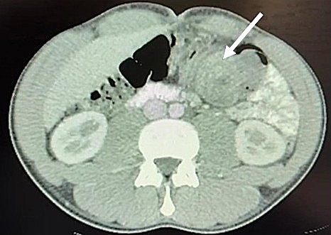
Abdominal ultrasonography revealed an intermittent “target sign” in the left flank, consistent with intussusception, associated with an irregularly contoured hypoechoic lesion measuring approximately 30 mm, suggestive of a lead point for the intussusception. Contrast-enhanced computed tomography (CT) of the chest, abdomen, and pelvis demonstrated an intraluminal lesion in the distal transverse colon associated with early intussusception, without evidence of perforation or free intraperitoneal fluid (Fig. 1).
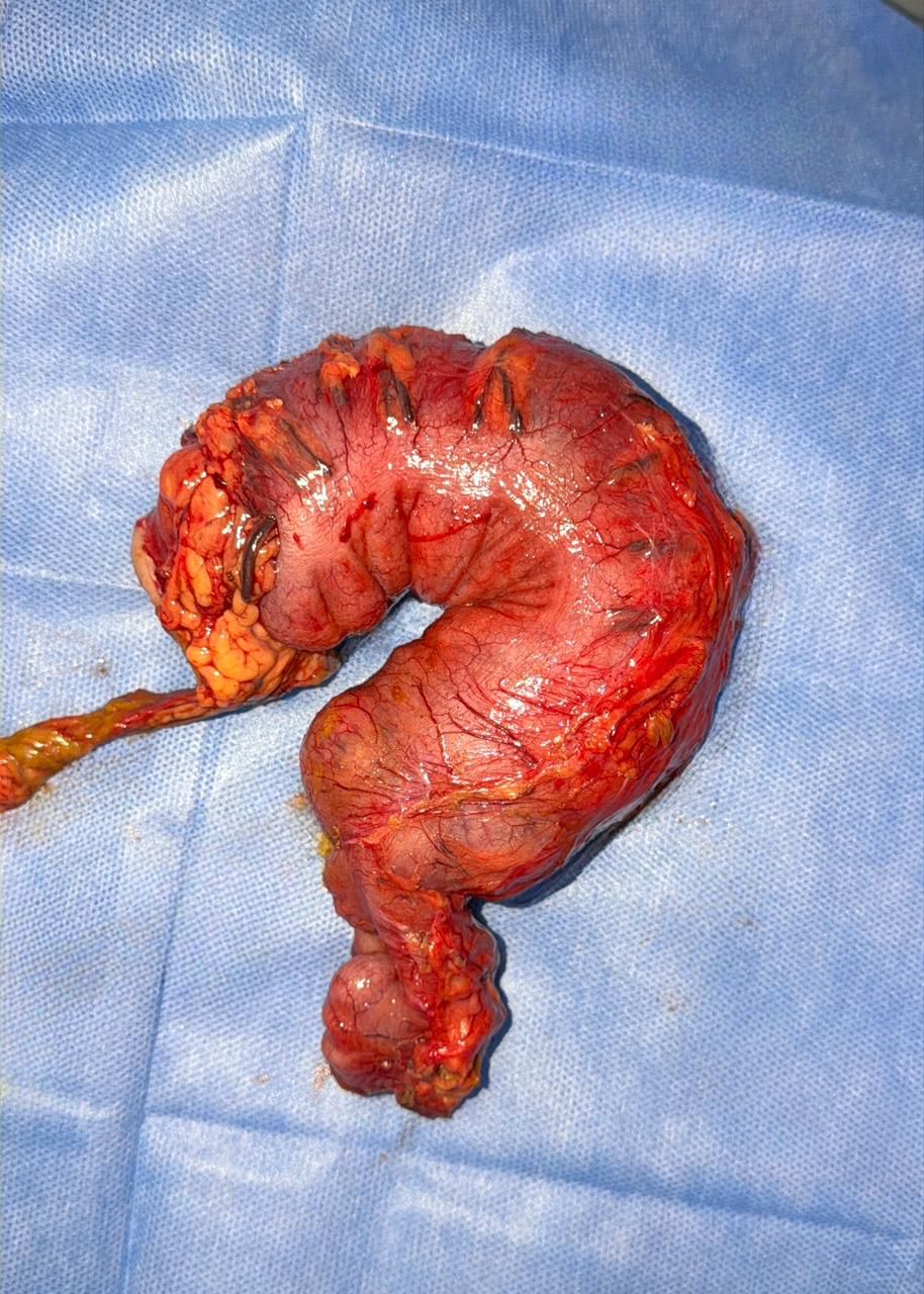
Exploratory laparotomy was performed, revealing a distal transverse colon tumor measuring approximately 5 cm in diameter. The lesion showed no serosal involvement but was firmly adherent to the deep planes, with proximal intussusception of the transverse colon. A segmental left colectomy with primary side-to-side anastomosis was performed (Fig. 2).
The postoperative course was uneventful, with progressive return of bowel function, adequate oral intake, and no complications. The patient was discharged on postoperative day 7.
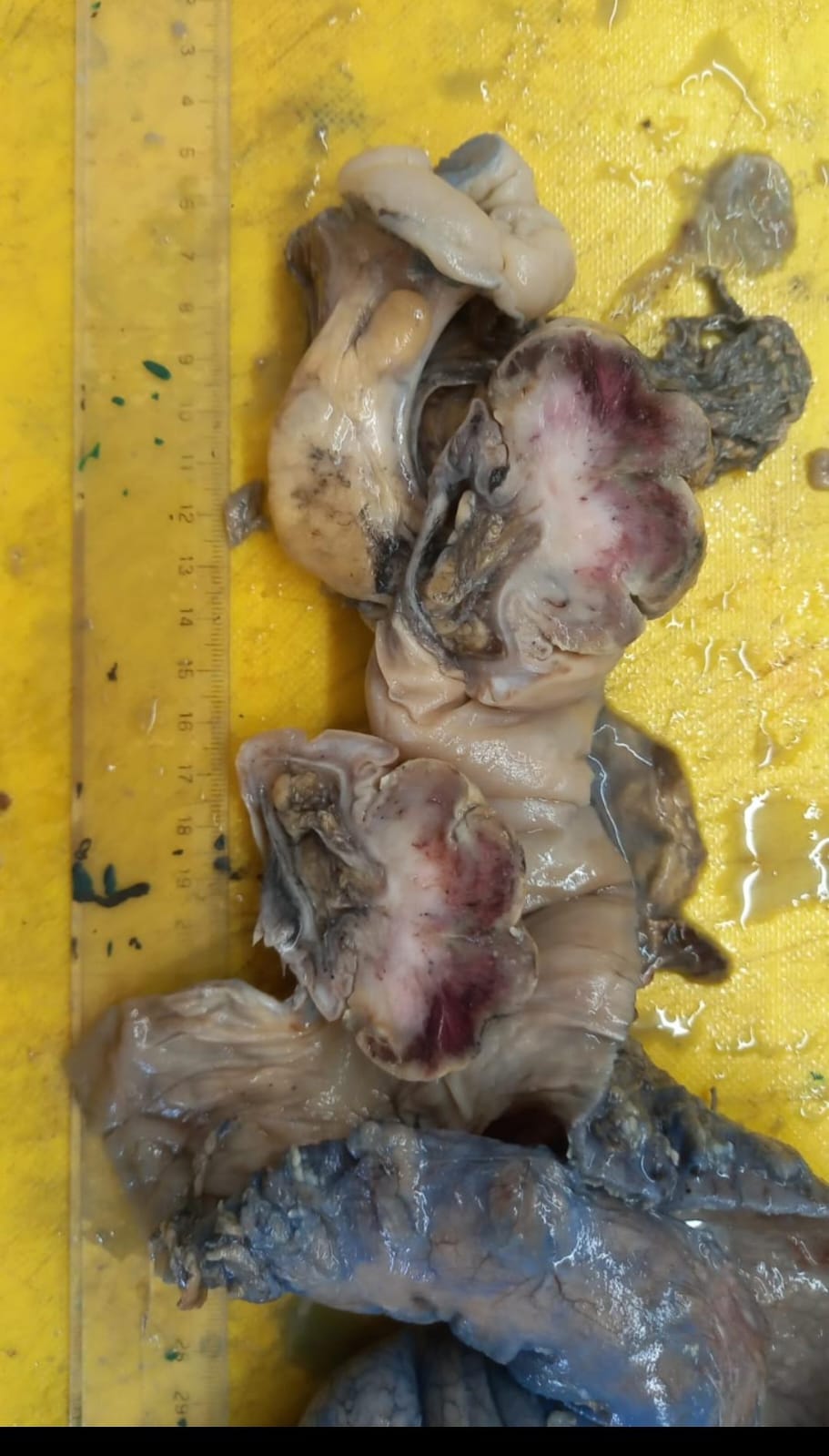
Gross pathological examination revealed intussusception at the proximal aspect of the surgical specimen. Upon opening the specimen, a mucosal tumor measuring 7.5 × 4.3 × 5 cm was identified, involving 50% of the circumference and causing complete luminal obstruction. The remaining mucosa showed loss of normal folds and an edematous appearance (Fig. 3).
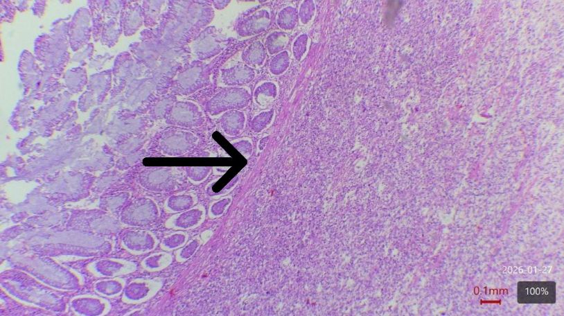
Histopathological examination revealed a proliferation of spindle cells with moderate cytologic atypia and transmural involvement.
The lesion was superficially ulcerated and covered by a fibrinoleukocytic pseudomembrane. Twelve lymph nodes were identified in the pericolonic adipose tissue, all showing reactive lymphadenitis (Fig. 4).
Immunohistochemical staining showed negative results for smooth muscle actin (SMA), S100 protein, CD117, DOG1, desmin, ALK, TLE1, and STAT6; patchy positivity for CD34; and a Ki-67 proliferation index of 20%. These findings supported the diagnosis of an intermediate-grade undifferentiated spindle cell sarcoma. The tumor was classified as FNCLCC (French Fédération Nationale des Centres de Lutte Contre le Cancer) grade 2, with a total score of 5 points based on differentiation score 3, mitotic count score 1, and necrosis score 1. This classification indicates intermediate biological aggressiveness and a moderate risk of recurrence and metastasis.
The patient remains under multidisciplinary follow-up with medical oncology, nutrition, and the colorectal surgery team.
The clinical presentation of gastrointestinal sarcomas is often nonspecific, contributing to delays in diagnosis. Patients typically present with abdominal pain, abdominal distention, altered bowel habits, a palpable abdominal mass, or symptoms of partial bowel obstruction; in some cases, the condition may progress to acute bowel obstruction.⁴ In the present case, the initial presentation was acute, with rapid symptom progression requiring urgent surgical intervention.
Diagnostic evaluation should be guided by the clinical presentation. Contrast-enhanced CT of the chest, abdomen, and pelvis is the primary imaging modality for diagnosis and staging, allowing assessment of tumor location, involvement of adjacent structures, and the presence of metastatic disease. Magnetic resonance imaging may be useful in selected cases, whereas positron emission tomography/computed tomography (PET/CT) is generally reserved for the characterization of indeterminate findings or the detection of distant disease.⁵
The role of colonoscopy in these tumors remains controversial. In early-stage lesions, the mucosa may remain intact, limiting endoscopic detection. However, when mucosal invasion or significant luminal involvement is present, colonoscopy enables direct tumor visualization and targeted biopsy acquisition.⁶
The management of soft tissue sarcomas is multimodal and requires a multidisciplinary approach. Surgery with curative intent remains the cornerstone of treatment. Nevertheless, local recurrence and metastatic disease may occur despite complete surgical resection. Undifferentiated spindle cell sarcoma predominantly spreads through the hematogenous route, with the lungs representing the most common site of metastasis, followed by the liver, bone, and peritoneum.3,5-7
Definitive diagnosis is established by histopathologic examination, with immunohistochemistry playing a central role, as the diagnosis is made by exclusion after specific lines of differentiation have been ruled out. Tumor grade should be reported in all cases because of its prognostic significance, most commonly using the FNCLCC grading system.⁵ Tumor staging is based on the American Joint Committee on Cancer (AJCC) TNM classification for soft tissue sarcomas (8th edition).⁸ In the present case, the tumor was classified as FNCLCC grade 2 and AJCC stage III (T2N0M0).
The rarity and histopathologic heterogeneity of soft tissue sarcomas pose substantial diagnostic challenges. A significant rate of diagnostic discordance has been reported between non-specialized institutions and referral centers, underscoring the importance of pathologic review by pathologists with expertise in soft tissue tumors.2,4
Available evidence suggests that primary colorectal sarcomas are characterized by aggressive biological behavior, with reported recurrence rates ranging from 20% to 85%. Median survival times have been reported to range from 30 to 53 months, reflecting their unfavorable prognosis.⁷
High-grade undifferentiated spindle cell sarcoma is an exceedingly rare neoplasm, with an exceptional location in the colon and significant diagnostic challenges, in which immunohistochemistry plays a pivotal role. Only a limited number of cases have been reported to date, and no controlled studies assessing the management of this entity are currently available. Further investigation of this rare tumor is warranted to establish standardized treatment strategies and multidisciplinary surveillance protocols in specialized centers, to improve long-term survival outcomes.
Author Contributions:
AVR: Conceptualization, methodology, formal analysis, and drafting of the original manuscript. CN: Data curation and formal analysis. FL: Supervision, methodology, and drafting – revision and editing. GRG: Supervision, drafting – revision and editing, and approval of the final version.
All authors approved the final version of the manuscript.
Conflict of interest statement: None.
Funding: None.
Data availability statement: The data are publicly available.
ORCIDs:
Andrea Veronica Ruchinsky: https://orcid.org/0009-0007-2325-4280
Camila J Navas: https://orcid.org/0009-0001-7778-6684
Florencia Litovska: https://orcid.org/0009-0009-7944-7924
Gabriela Rio Gorrini: https://orcid.org/0009-0006-2335-5471
Nagtegaal ID, Odze RD, Klimstra D, Paradis V, Rugge M, Schirmacher P, et al. The 2019 WHO classification of tumours of the digestive system. Histopathology. 2020;76(2):182–8. https://doi.org/10.1111/his.13975
Sbaraglia M, Bellan E, Dei Tos AP. (2021). The 2020 WHO classification of soft tissue tumours: news and perspectives. Pathologica.113(2);70–84. https://doi.org/10.32074/1591-951X-213
Qureshi PAAA, Zeb S, Tryggvason T, Þórisson A. Undifferentiated pleomorphic sarcoma of the descending colon: an infrequent occurrence. Cureus. 2024;16(5):e61346. doi:10.7759/cureus.61346. PMID:38947723.
Wu L, Wu H, Huang F, Li X, Zhen Y, Cheng H, et al. Primary undifferentiated sarcoma of the transverse colon in a young female patient: case report. Research Square. [Internet]. 2023 Jun 28 [cited 2026 Jan 26]. Available from: https://doi.org/10.21203/rs.3.rs-3094909/v1
Gronchi A, Miah AB, Dei Tos AP, Abecassis N, Bajpai J, Bauer S, et al. Soft tissue and visceral sarcomas: ESMO–EURACAN–GENTURIS Clinical Practice Guidelines for diagnosis, treatment and follow-up. Ann Oncol. 2021;32(11):1348–65. doi:10.1016/j.annonc.2021.07.006.
Han X, Zhao L, Mu Y, Liu G., Zhao G, He H, et al. Undifferentiated high-grade pleomorphic sarcoma of the colon: a rare case report and literature review. BMC Gastroenterol. 2022; 22(1):115. https://doi.org/10.1186/s12876-022-02189-x
Thiels CA, Bergquist JR, Krajewski AC, Lee HE, Nelson H, Mathis KL, et al. Outcomes of primary colorectal sarcoma: a National Cancer Data Base (NCDB) review. J Gastrointest Surg. 2017;21(3):560–8. https://doi.org/10.1007/s11605-016-3347-0
Amin MB, Edge SB, Greene FL, Byrd DR, Brookland RK, Washington MK, et al., eds. AJCC Cancer Staging Manual. 8th ed. New York: Springer; 2017.